Dermal fillers are among the most commonly performed cosmetic procedures worldwide, with over 6 million sessions annually in the United States alone. Most go smoothly. But when filler material is accidentally injected into a blood vessel that connects to the ophthalmic artery — the artery that supplies the eye — the result can be sudden, irreversible blindness.
The risk is not theoretical. Early reviews documented 98 published cases by 2015, with an additional 48 new cases added by 2019, and the American Academy of Ophthalmology noted 190 reported cases from 1906 through 2019. A comprehensive 2024 update by Doyon et al. identified 365 additional cases published between 2018 and 2023, bringing the cumulative total to over 500 published cases. Because reporting is voluntary and inconsistent, the true number is almost certainly higher.
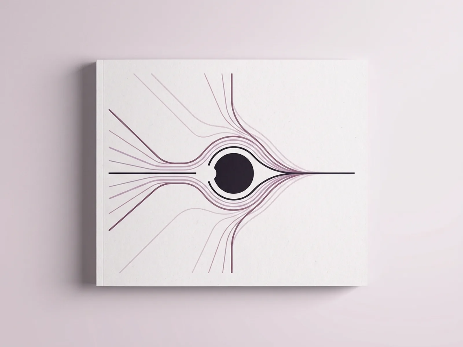
This article explains the mechanism behind filler-induced blindness, which facial areas carry the greatest danger, what symptoms indicate an ocular emergency, what rescue attempts exist (and their limitations), and what every patient should ask before agreeing to facial filler.
How filler causes blindness
Blindness from filler occurs through a process called retrograde intra-arterial embolism. Three conditions must be present simultaneously:
Injection pressure exceeds systolic blood pressure. The average pressure required to embolize the ophthalmic artery is approximately 166.7 mmHg. Studies show that it is easy for an injector to generate pressures well above 200 mmHg within 2 to 3 seconds of pushing the syringe plunger.
The needle or cannula tip is inside the lumen of an artery that connects to the ophthalmic artery — typically the supratrochlear, supraorbital, or dorsal nasal artery.
Sufficient filler volume enters the vessel. The entire volume of the supratrochlear artery from the glabella to the orbital apex is only about 0.085 mL (range 0.04–0.12 mL). Even very small injected volumes can exceed this capacity.
When these conditions align, filler material is forced backward (retrograde) through the artery toward the ophthalmic artery and then forward (anterograde) into the central retinal artery or its branches, blocking blood supply to the retina. The retina is extraordinarily sensitive to oxygen deprivation — irreversible damage begins within 12 to 15 minutes of complete occlusion.
The danger zones
Not all facial areas carry equal risk. The nasal region has now overtaken the glabella as the highest-risk site for filler-induced vision loss, according to a 2020 consensus.
Highest-risk areas:
- Nose (nonsurgical rhinoplasty, nasal dorsum, nasal tip). The dorsal nasal artery and angular artery anastomose directly with the ophthalmic artery. In a 2024 case series published in Scientific Reports, the nose was the most common injection site associated with catastrophic ocular complications (50% of cases).
- Glabella (between the eyebrows). The supratrochlear and supraorbital arteries supply this area and are direct branches of the ophthalmic artery.
- Forehead. The supraorbital and supratrochlear arteries traverse this region.
- Periorbital area (around the eyes). Including tear troughs and the temples, where the superficial temporal artery and other orbital branches are present.
Moderate-risk areas:
- Nasolabial folds (angular artery branches)
- Mid-face and cheeks (when injected superficially near vascular pathways)
Both sharp needles and blunt cannulas have been associated with cases of blindness. Neither tool eliminates the risk.
Symptoms: this is an ocular emergency
Blindness from filler typically presents during or immediately after injection. The onset is usually sudden. Key symptoms:
- Sudden vision loss in one or both eyes — most cases present as total blindness within minutes
- Severe eye pain
- Ptosis (drooping eyelid)
- Ophthalmoplegia (inability to move the eye)
- Headache, nausea, or vomiting
- In some cases: stroke symptoms including weakness, hemiplegia, or altered mental status
No light perception is reported in up to 68% of cases. If any of these symptoms occur during or after a filler injection, the patient should be transferred immediately to the nearest emergency department with ophthalmology and stroke capability — by ambulance, not by private car.
Rescue attempts and why prevention matters most
There is no proven treatment for filler-induced blindness. Several interventions are attempted, but none has demonstrated consistent success.
Hyaluronidase
For HA fillers, hyaluronidase — the enzyme that dissolves hyaluronic acid — is the primary tool. It can be administered several ways:
- Periocular (around the eye) injection. Subcutaneous hyaluronidase near the affected area. A systematic review found hyaluronidase was used in 86% of vision loss cases but was successful in only 11.1%.
- Retrobulbar injection. Hyaluronidase (typically 1,500 IU in 4 mL saline) injected behind the eye using a 25G needle. Doyon et al. (2024) found this improved vision in only 2 of 38 patients (5.3%). The American Academy of Ophthalmology notes there is no well-documented case of vision returning from a confirmed embolism after retrobulbar hyaluronidase.
- Intra-arterial hyaluronidase. Delivered via catheterization of the ophthalmic artery. A 2024 Scientific Reports case series found this alleviated occlusion in 5 patients but resulted in significant visual improvement in only 2.
Supportive measures
- Ocular massage to lower intraocular pressure and potentially dislodge the embolus
- Anterior chamber paracentesis (draining fluid from the eye)
- Rebreathing into a paper bag (to raise blood CO₂ and dilate vessels)
- Sublingual glycerol trinitrate for vasodilation
The RANZCO (Royal Australian and New Zealand College of Ophthalmologists) 2024 guideline recommends that injectors accompany the patient to the emergency department, bring hyaluronidase with them, and perform serial vision and neurological assessments during transfer.
The critical time window
Animal studies show that restoring blood flow within 90 minutes may allow some retinal recovery. After 100 minutes, outcomes become variable. After 4 hours of non-perfusion, retinal infarction is profound and irreversible. This is why immediate transfer to a stroke-capable center is essential.
Why outcomes are generally poor
Several factors make filler-induced blindness exceptionally difficult to reverse:
- Speed of onset. Occlusion is often complete within seconds, and the retina begins dying within minutes.
- Physical properties of HA. Highly cross-linked HA fillers may resist breakdown by hyaluronidase, especially when trapped in the retinal microvasculature.
- Injection pressure. By the time symptoms appear, the damage cascade is already underway.
- Delay to treatment. Many patients are in outpatient clinics without immediate ophthalmology access.
Risk factors that increase danger
- Injection technique. High-pressure bolus injection into danger zones is the primary risk factor. Slow, low-volume injection with frequent aspiration reduces — but does not eliminate — risk.
- Filler type. All fillers can cause blindness, but autologous fat carries the highest rate of complete vision loss (80% of cases vs. 50% for HA), because fat particles cannot be dissolved. HA fillers, while still dangerous, at least offer the theoretical possibility of enzymatic dissolution.
- Lidocaine in filler. Many HA fillers contain lidocaine, which can mask the immediate pain of intravascular injection, delaying recognition.
- Provider anatomy knowledge. Injectors without detailed training in facial vascular anatomy are more likely to inadvertently cannulate dangerous vessels.
- Prior surgery or filler. Altered anatomy from previous procedures can shift vessel positions.
What to ask before a filler appointment
- "What is the risk of blindness from this procedure?" Any injector who dismisses or minimizes this risk is not providing adequate informed consent. Vision loss should be discussed as part of the consent process.
- "Which areas are being treated, and what is their risk level?" Nose, glabella, forehead, and periorbital areas are highest risk. If you are being treated in these zones, ask specifically what precautions the provider takes.
- "Do you have hyaluronidase on-site and an emergency protocol?" The provider should be able to describe a written emergency plan for ocular complications, including immediate referral to ophthalmology.
- "What technique do you use in danger zones?" Aspiration before injection, slow low-volume delivery, deep periosteal placement where appropriate, and avoidance of bolus technique in high-risk areas are all standard safety measures.
- "What warning signs should I watch for?" You should receive written instructions describing vision changes, eye pain, ptosis, and skin color changes — with a 24/7 contact number.
- "What are your credentials and training?" Board-certified dermatologists, plastic surgeons, and oculoplastic surgeons have the deepest training in facial vascular anatomy and complication management.
The bottom line
Filler-induced blindness is rare but catastrophic. There is no reliable treatment once it occurs — outcomes are poor even with aggressive intervention. Prevention through careful technique, thorough knowledge of vascular anatomy, and informed patient consent is the only meaningful strategy. Patients considering filler in the nose, glabella, forehead, or periorbital area should understand the risk, confirm their provider has an emergency protocol, and know the warning signs that demand immediate emergency evaluation.
Sources
- Beleznay K, Carruthers JDA, Humphrey S, Jones D. "Avoiding and Treating Blindness From Fillers: A Review of the World Literature." Dermatol Surg. 2015;41(10):1097–1117. https://pubmed.ncbi.nlm.nih.gov/26355853/
- Beleznay K, et al. "Update on Avoiding and Treating Blindness From Fillers: A Recent Review of the World Literature." Aesthet Surg J. 2019;39(6):662–674. https://academic.oup.com/asj/article/39/6/662/5414135
- Doyon VC, Liu C, Fitzgerald R, et al. "Update on Blindness From Filler: Review of Prognostic Factors, Management Approaches, and a Century of Published Cases." Aesthet Surg J. 2024;44(10):1091–1104. https://academic.oup.com/asj/article/44/10/1091/7682938
- Zhang L, et al. "Disastrous cerebral and ocular vascular complications after cosmetic facial filler injections." Scientific Reports. 2024. https://www.nature.com/articles/s41598-024-54202-w
- RANZCO. "Guideline for Filler Blindness." 2024. https://ranzco.edu/wp-content/uploads/2024/06/RANZCO-Filler-Blindness-Guidelines_2024.pdf
- American Academy of Ophthalmology. "Safety Update: Periocular Dermal Fillers." EyeNet Magazine. https://www.aao.org/eyenet/article/safety-update-periocular-dermal-fillers
- U.S. Food and Drug Administration. "Dermal Fillers — Approved Dermal Fillers." https://www.fda.gov/medical-devices/cosmetic-devices/dermal-fillers
- Goodman G, et al. "A Consensus on Minimizing the Risk of Hyaluronic Acid Embolic Visual Loss." Aesthet Surg J. 2020;40:1009–1021. https://academic.oup.com/asj/article/40/9/1009/5845299
- Murthy R, Roos J, Goldberg R. "Periocular hyaluronic acid fillers: applications, implications, complications." Curr Opin Ophthalmol. 2019;30(5):395–400. https://pubmed.ncbi.nlm.nih.gov/31343436/
- Hwang CJ, et al. "Blindness after filler injection: mechanism and treatment." Facial Plast Surg Clin North Am. 2021. https://pubmed.ncbi.nlm.nih.gov/33962357/