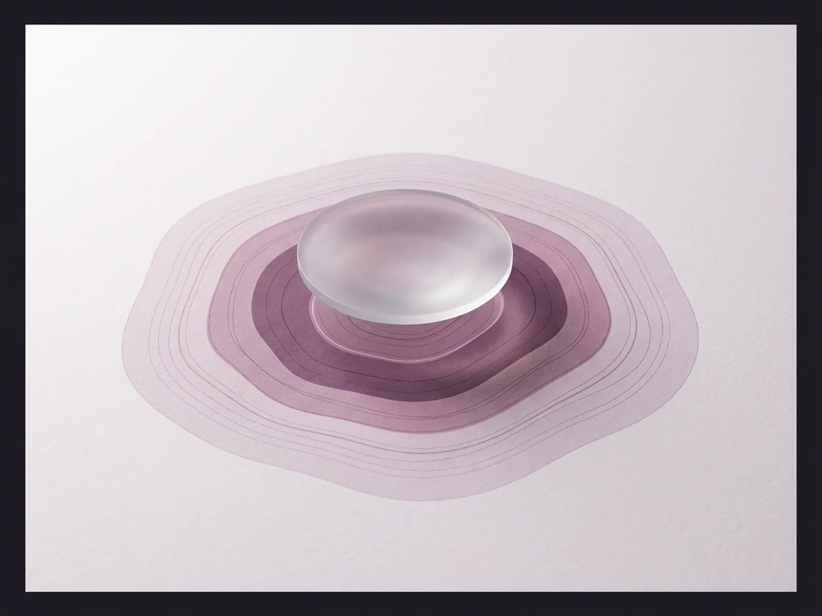
Filler migration means that an injected dermal filler has moved from its intended placement site into adjacent tissue. It is not the same as post-injection swelling, which is expected and resolves within days. Migration is a structural event: the product has physically relocated, and the result is a visible change in contour that does not improve on its own.
The concept entered public conversation largely through lip filler. Patients who had their vermilion border injected to enhance lip shape sometimes noticed, months or years later, a visible ridge of product above the lip line — a "shelf" or "duck lip" appearance that was not there immediately after treatment. But migration is not limited to lips. It also occurs in the tear troughs, cheeks, jawline, and nasolabial folds, and it can involve any injectable filler, not just hyaluronic acid.
Understanding what migration is — and what it is not —requires separating three phenomena that patients often conflate: actual product displacement, water retention from hydrophilic filler, and age-related volume changes that were previously masked by filler.
What migration actually looks like
Actual filler migration produces a contour change in a location different from where the product was injected. The most common presentation is a palpable and visible ridge of material above the vermilion border of the lip. In the tear troughs, migrated filler can create a bulge visible in the lower eyelid or cheek. In the jawline, it can produce a blunted or irregular contour.
Patients often search for "did my filler migrate?" when they notice changes that may have simpler explanations:
- Post-injection edema. Hyaluronic acid fillers are hydrophilic — they attract and bind water. After injection, the local swelling this causes can look like migration, especially in thin-skinned areas like the tear troughs. This swelling typically peaks at 24 to 72 hours and resolves over one to two weeks. It is not migration.
- Water retention from fraying HA chains. Over time, the cross-linked HA in filler begins to break down. As the polymer chains fray, they expose more binding sites for water molecules. This can cause a gradual increase in apparent volume that looks like the filler has spread, but the product has not actually moved. It is attracting more moisture than it did when freshly injected.
- Natural aging and tissue laxity. Filler masks volume loss. As the face continues to age, the tissue around the filler changes. What looks like migration can sometimes be the face settling around a static filler deposit — the product did not move, but the surrounding anatomy shifted.
Actual migration is confirmed when the product can be identified in a location distinct from its injection site, either by clinical examination, ultrasound, or MRI. This distinction matters because the treatment for each scenario is different.
Why filler migrates
Migration has multiple contributing causes, and in most clinical cases, more than one factor is involved.
Excessive volume. Injecting more filler than the tissue compartment can hold increases the pressure gradient that pushes product into surrounding tissue. This is the most commonly cited cause in the clinical literature. The tissue has a finite capacity; exceeding it forces material outward along planes of least resistance.
Incorrect tissue plane. Fillers are designed for specific depths. Superficial placement of a product intended for deep injection can allow it to move more freely through tissue. Similarly, placing a volumizing filler in the superficial fat pad rather than the deep fat compartment increases the chance of visible displacement.
High-pressure injection. Rapid injection under high pressure forces filler into a wider area than intended. A controlled, slow injection allows the product to deposit cleanly within the target compartment.
Product choice. Softer, more fluid fillers with lower G-prime (the measure of gel firmness) are more prone to spreading within tissue than firmer, more cohesive products. A filler designed for lip body may not hold its shape when used at the vermilion border, and vice versa.
Repeated treatments without accounting for residual product. Patients who receive filler in the same area every few months may have significant residual product that was not fully metabolized. Adding more filler on top of existing product increases total volume and the risk of displacement. Many injectors do not assess for residual product before re-treating.
Anatomical movement. The lips are the clearest example. The orbicularis oris muscle is in near-constant motion during speech, eating, and facial expression. This mechanical activity acts on the filler over time, gradually pushing it outward. Areas with less muscular activity are less susceptible to this mechanism.
Which areas are most prone
Lips are the most commonly discussed site for migration, and the anatomy explains why. The vermilion border is a thin, mobile structure reinforced by the orbicularis oris. Filler placed at or near the border is subjected to thousands of compressive cycles per day. Migration above the border into the white lip creates the characteristic ridge that patients describe as "filler lips" or "duck lips."
Tear troughs are the second most common site. The thin skin in this area makes any volume change visible. Filler placed too superficially or in excess can spread superiorly into the lower eyelid or inferiorly into the cheek, creating visible bulges.
Cheeks can experience migration when large volumes of volumizing filler are placed without adequate compartmental control. Product placed in the deep medial cheek fat pad can extend into the suborbital area, creating lower eyelid fullness that was not intended.
Jawline and nasolabial folds are less commonly affected but not immune. Migration in these areas typically results from overfilling rather than mechanical displacement.
HA vs non-HA filler migration: a critical distinction
Hyaluronic acid fillers (Juvéderm, Restylane, Belotero, RHA) can be dissolved with hyaluronidase. This makes migration of HA fillers a reversible problem in most cases.
Non-HA fillers cannot be dissolved. This includes:
- Calcium hydroxylapatite (Radiesse): A mineral-based filler that stimulates collagen production. Migration of Radiesse is rare because the product is thick and typically placed deep, but if it does move, there is no enzyme that can reverse it.
- Poly-L-lactic acid (Sculptra): A biostimulatory filler that works by triggering collagen synthesis over months. Sculptra particles are not a gel — they are suspended in a carrier fluid that is absorbed within days. True migration of Sculptra particles is uncommon, but if it occurs, the body must metabolize the particles over its natural timeline, which can take one to two years.
- PMMA (Bellafill): A permanent filler containing polymethylmethacrylate microspheres. Migration of PMMA is irreversible without surgical excision.
This distinction is one of the strongest arguments for using HA fillers in high-mobility areas like the lips, where migration risk is highest. If a permanent or semi-permanent filler migrates in a visible area, the patient has very few options.
The imaging evidence
For years, filler migration was discussed primarily as a clinical observation — injectors and patients noticed contour changes that suggested product had moved, but imaging confirmation was limited.
That changed when a surgeon's video showing MRI scans of a patient who had received 12 syringes of HA filler over six years went viral. The MRI showed visible filler material distributed well beyond the original injection sites. The case was extreme — 12 syringes is a large cumulative volume — but it provided the first widespread public visual evidence that filler does not always stay where it is placed.
The formal research is now catching up. ClinicalTrials.gov NCT07079397 is an ongoing study sponsored by Galderma titled "The Integration Pattern of NASHA and OBT-Based Fillers: An Ultrasound and MRI Study." The study, which began in December 2025 and is expected to complete in 2027, uses both ultrasound and MRI to evaluate how different Galderma filler formulations (NASHA-based products like Restylane and OBT-based products like Restylane Lyft) integrate into tissue over time — and whether they migrate. This is the first formal prospective clinical trial designed specifically to study filler migration using advanced imaging.
The distinction between NASHA (Non-Animal Stabilized Hyaluronic Acid) and OBT (Optimized Balance Technology) matters because these represent different gel architectures with different tissue-integration profiles. Whether one is more prone to migration than the other has been debated anecdotally; this study aims to answer the question with controlled data.
Until those results are published, the clinical literature on migration relies on retrospective analysis and case series. A 2025 review in PMC (PMC12011065) on the management of delayed complications of HA fillers identified filler migration as one of several late-onset adverse events, alongside foreign body granuloma reactions and delayed hypersensitivity. The authors noted that late complications are likely under-reported because many patients do not connect contour changes months or years later to a prior injection.
Treatment: when and how to address migration
For HA filler migration, hyaluronidase is the primary treatment. Hyaluronidase is an enzyme that breaks down hyaluronic acid by cleaving its glycosidic bonds. Injected into or near the migrated product, it begins working within minutes and produces visible reduction within 24 to 48 hours.
But hyaluronidase is not perfectly selective. It dissolves all hyaluronic acid it encounters, including the HA that naturally exists in skin, connective tissue, and joint fluid. This means the treatment has trade-offs:
- The minimum effective dose should be used. Over-injecting hyaluronidase can degrade the patient's own tissue HA, leaving the area temporarily deflated, dry, or hollow.
- Staged sessions are often preferable to a single aggressive treatment. Waiting at least a week between sessions allows the provider to assess how much product was dissolved and whether the contour has improved, before deciding whether more is needed.
- Ultrasound guidance improves precision. A 2024 study by Bravo et al. in the Journal of Cosmetic Dermatology investigated ultrasound-guided hyaluronidase injection, concluding that real-time imaging allows the provider to place the enzyme directly into the migrated deposit rather than distributing it through surrounding tissue. This reduces the total dose required and limits collateral damage to natural HA.
A 2025 study by Hong et al. in the Journal of Cosmetic Dermatology reviewed hyaluronidase use protocols and emphasized that dosing should be calibrated to the volume and age of the filler deposit, not applied at a fixed standard dose.
Patients should also be aware of what Wilde et al. termed "posthyaluronidase syndrome" in a 2024 paper in Plastic and Reconstructive Surgery. The authors identified predictors of poor outcomes after hyaluronidase treatment, including patients who had large volumes of long-standing filler dissolved in a single session. The lesson: aggressive dissolution of years of accumulated filler can produce aesthetically and emotionally distressing results. A gradual approach is safer.
For non-HA filler migration, options are limited. The body metabolizes calcium hydroxylapatite over 12 to 18 months and poly-L-lactic acid over one to two years. If the patient is willing to wait, the product will eventually be absorbed. If the migration is causing significant aesthetic or functional problems, surgical excision may be considered, but this is a last resort with its own risks including scarring and nerve damage.
UPMC HealthBeat notes that HA filler dissolves naturally in 6 to 12 months without intervention. For patients with mild migration who are not in a hurry, waiting is a reasonable option.
Prevention: what to ask before treatment
Migration is far easier to prevent than to treat. Before agreeing to filler, patients should have a direct conversation with their injector that covers:
- How much volume are you planning, and is it conservative? More is not better. The lowest effective volume is safest.
- What product are you using, and is it appropriate for this area? Lip border filler and cheek volumizer are different products. Ask whether the specific formulation matches the anatomical site.
- Will you assess for residual filler before adding more? If you have been treated before, the injector should evaluate what remains before injecting additional product.
- What tissue plane are you targeting? A knowledgeable injector can explain whether the product is going into superficial or deep tissue and why.
- What is the injection technique? Slow, low-pressure injection with a cannula (rather than a sharp needle) can reduce the risk of product spreading beyond the target area.
- What are the aftercare instructions? Avoiding pressure, massage, and extreme facial movement for the first 24 to 48 hours after lip filler gives the product time to integrate into the tissue before mechanical forces act on it.
- What is the plan if the result is not right? An injector who is confident in their technique should be willing to discuss revision options, including hyaluronidase, before the procedure — not only after something goes wrong.
The single most effective prevention strategy is choosing an experienced, board-certified injector who works with the full range of filler products and has the anatomical knowledge to place product correctly the first time. Migration is, in most cases, a technique-sensitive problem. The provider matters more than the product.
Sources
- Management of delayed complications of hyaluronic acid fillers. PMC. 2025. https://pmc.ncbi.nlm.nih.gov/articles/PMC12011065/
- The Integration Pattern of NASHA and OBT-Based Fillers: An Ultrasound and MRI Study. ClinicalTrials.gov NCT07079397. Galderma. 2025–2027. https://clinicaltrials.gov/study/NCT07079397
- Hong et al. Hyaluronidase use protocols for HA filler complications. Journal of Cosmetic Dermatology. 2025.
- Wilde et al. Predictors of poor outcomes after hyaluronidase: "posthyaluronidase syndrome." Plastic and Reconstructive Surgery. 2024.
- Bravo et al. Ultrasound-guided hyaluronidase investigation for filler complications. Journal of Cosmetic Dermatology. 2024.
- Guideline for the safe use of hyaluronidase in aesthetic medicine. PMC. 2021. https://pmc.ncbi.nlm.nih.gov/articles/PMC8570661/
- How long does dermal filler last? UPMC HealthBeat. https://www.upmc.com/services/skin-services/resources/how-long-does-dermal-filler-last
- Risks and rewards: what to know about dissolving filler. American Society of Plastic Surgeons. https://www.plasticsurgery.org/news/articles/risks-and-rewards-what-to-know-about-dissolving-filler
- FDA Executive Summary: General Issues Panel Meeting on Dermal Fillers. August 2025. https://www.fda.gov/media/188185/download