Dermal fillers are the second most common minimally invasive cosmetic procedure in the United States, with an estimated 6.26 million procedures performed in 2024. Most sessions are uneventful. But when filler material enters a blood vessel or compresses it from outside, the resulting vascular occlusion can cut off blood supply to surrounding tissue — and in the worst cases, to the eye or brain.
Vascular occlusion is rare but not theoretical. The FDA's Manufacturer and User Facility Device Experience (MAUDE) database has logged 17,768 serious injury reports related to dermal fillers, with vascular system impairment, obstruction, and occlusion among the most commonly reported serious adverse events. A 2025 study of 100 filler-related vascular complications across six international centers, presented at the Radiological Society of North America, found that in one-third of cases blood flow was absent in major facial blood vessels.
This article explains what vascular occlusion is, which facial areas carry the highest risk, how to tell the difference between normal post-injection bruising and a vascular event, what the emergency protocol looks like, and what every patient should ask before a filler appointment.
What vascular occlusion is
Vascular occlusion occurs when dermal filler material either enters a blood vessel directly (intravascular embolism) or compresses a vessel from the outside (extravascular compression). Both scenarios reduce or block blood flow to downstream tissue.
When an artery is blocked, the tissue it supplies is deprived of oxygen. Without intervention, this leads to ischemia (insufficient blood flow), progressing to tissue death (necrosis) within hours to days. If the blocked vessel communicates with the ophthalmic artery — which supplies the eye — the result can be permanent vision loss or blindness.
Any injectable filler can cause vascular occlusion. Hyaluronic acid (HA) fillers, which represent approximately 80% of the dermal filler market, are the most frequently implicated simply because they are the most commonly used. But non-HA fillers — including calcium hydroxylapatite (Radiesse), poly-L-lactic acid (Sculptra), and polymethylmethacrylate (Bellafill) — carry the same fundamental risk.
How common it is
The incidence of vascular occlusion is difficult to pin down precisely because reporting is inconsistent and many mild cases resolve before diagnosis. Available estimates:
- A 2013 analysis estimated the incidence of impending skin necrosis following filler treatment at approximately 0.001% (1 in 100,000 cases).
- By 2020, this estimate had risen to 0.009% — a ninefold increase, likely reflecting both higher procedure volumes and improved recognition and reporting.
- The FDA MAUDE database shows a steady increase in serious injury reports year over year, with 1,478 reports in 2023 and 1,179 through November 2024.
The numbers are low relative to the millions of filler procedures performed annually. But the consequences of a missed or delayed diagnosis — scarring, disfigurement, permanent vision loss — are severe enough that both patients and providers should be prepared.
High-risk facial zones
The face has an extensive network of blood vessels, and some areas are more dangerous than others for filler injection.
Highest-risk areas:
- Nose and nasal alar region. The angular artery and dorsal nasal artery communicate with the ophthalmic artery system. Fillers injected into or around the nose (including "liquid rhinoplasty") carry the highest risk of blindness.
- Glabella (between the eyebrows). The supratrochlear and supraorbital arteries supply this area and connect directly to the ophthalmic artery.
- Forehead. The supraorbital and supratrochlear arteries run through this region.
- Nasolabial folds. The facial artery and its branches (including the angular artery) traverse this zone.
Moderate-risk areas:
- Perioral region (lips, chin, oral commissures)
- Temples
Lower-risk areas (but not risk-free):
- Cheeks (when injected deeply, above the periosteum, away from major vessels)
- Jawline
A 2024 study published in Scientific Reports documented catastrophic cerebral and ocular complications — including blindness and cerebral infarction — following cosmetic facial filler injections, emphasizing that severe outcomes, while rare, do occur.
Signs of vascular occlusion: what to watch for
Vascular occlusion can present during injection, within minutes, or hours later. The signs are progressive — early recognition and immediate action are critical.
During or immediately after injection
- Sudden blanching or whitening of the skin in the distribution of the affected vessel
- Severe, sharp pain that is disproportionate to the injection itself — though this can be masked by lidocaine mixed into many fillers, delaying recognition
- Livedo or mottled discoloration — a reticulated (net-like) red or purple pattern
- Resistance or back-pressure on the syringe plunger (felt by the injector)
Important: Many modern HA fillers contain lidocaine (a local anesthetic). While this improves patient comfort, it can mask the early pain of vascular occlusion for several hours until the anesthetic wears off. This means that vascular occlusion is often not detected during the injection itself — it may be discovered only when the patient reports persistent pain, swelling, or color changes the next day. The ASDS task force emphasizes that all office staff, not just the injector, should be trained to recognize these symptoms when patients call with post-treatment concerns.
Minutes to hours after injection
- Dusky, bluish, or grayish skin tone in the affected area
- Delayed capillary refill — pressing the skin and releasing should return color within 1–2 seconds; in occlusion, color return is sluggish or absent
- Cool or pale skin compared to surrounding areas
- Increasing pain rather than the gradual improvement expected after filler
Hours to days (late signs)
- Skin breakdown, blistering, or crusting indicating tissue necrosis
- Darkening to purple or black — tissue death is progressing
- Eschar formation (dry, dark scab)
Ocular emergency signs
Any of these symptoms during or after a facial filler injection require immediate emergency referral to an ophthalmologist and emergency department:
- Sudden vision loss or changes in vision
- Eye pain
- Drooping eyelid (ptosis)
- Double vision
- Inability to move the eye normally
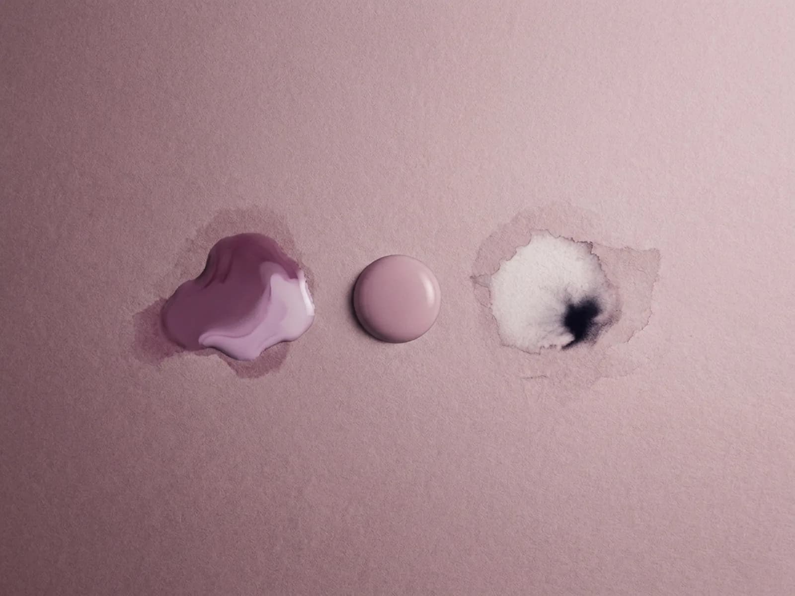
How to tell vascular occlusion from a normal bruise
Patients sometimes confuse post-injection bruising with vascular occlusion. The differences matter:
| Feature | Normal bruise | Vascular occlusion |
|---|---|---|
| Color | Reddish-purple, fades to yellow over days | Pale initially, then darkens to purple or black |
| Pain | Mild soreness, improving | Severe, worsening |
| Capillary refill | Normal (under 2 seconds) | Delayed or absent |
| Skin temperature | Normal | Cool |
| Progression | Improves over days | Worsens over hours |
If there is any doubt, the safest approach is to contact the injector immediately or go to an emergency department. Waiting to see if it improves on its own is not appropriate when vascular occlusion is possible.
How providers treat vascular occlusion
Hyaluronidase for HA fillers
For hyaluronic acid fillers, the primary emergency treatment is hyaluronidase — an enzyme that breaks down hyaluronic acid. The current consensus approach involves:
- High-dose pulsed hyaluronidase protocol (DeLorenzi): 450–1,500 IU total, injected perilesionally in up to 4 cycles. The goal is to flood the ischemic tissue with enough enzyme to dissolve the occluding filler and restore blood flow.
- Ultrasound-guided hyaluronidase: Using high-frequency ultrasound (HFUS) to visualize the exact location of the filler, allowing targeted injection with lower doses (35–50 IU). This approach requires specialized equipment and training.
- Intra-arterial hyaluronidase: Cannulating the facial artery to deliver enzyme directly into the vascular supply. This technique has shown promise in case reports but requires specialized interventional training.
A 2024 review in JMIR Dermatology emphasized that the success of hyaluronidase depends on timing, the specific HA filler's cross-linking density, and the size of the ischemic area. Animal studies show that administration within 4 hours of occlusion significantly improves outcomes.
For non-HA fillers
Non-HA fillers (Radiesse, Sculptra, Bellafill) cannot be dissolved with hyaluronidase. Management relies on:
- Supportive measures: warm compresses, aspirin, topical nitroglycerin paste to improve local blood flow
- Hyperbaric oxygen therapy (HBOT) in some cases
- Waiting for natural resorption of the material
This makes prevention and proper injection technique even more critical with non-HA fillers.
Additional treatments
The 2025 THIS and FAT protocol published in Frontiers in Medicine combines hyaluronidase with a nanofat membrane technique to support tissue regeneration after vascular occlusion. While promising, this approach requires specialized training and is not yet widely available.
What increases the risk
Several factors raise the probability of vascular occlusion:
- Injection technique. Bolus injection (pushing a large volume at once) carries higher risk than slow, small-volume delivery. Needle injection has a higher intravascular injection risk than blunt cannula in some (but not all) anatomical areas.
- Anatomical knowledge. Providers who lack detailed understanding of facial vascular anatomy — particularly the connections between facial arteries and the ophthalmic artery — are more likely to inject into or near dangerous vessels.
- High-viscosity fillers. Fillers with higher viscosity and greater cross-linking may form larger, more obstructive emboli if injected intravascularly.
- Volume injected. Larger volumes per injection point increase the risk of both intravascular injection and extravascular compression.
- Previous filler or surgery. Scarring and altered anatomy from prior procedures can shift the position of blood vessels, making them harder to avoid.
What to ask your injector
Before any filler appointment, ask these questions directly:
- What is your protocol if vascular occlusion occurs? A prepared provider should be able to describe their emergency plan without hesitation.
- Do you have hyaluronidase on-site? If you are receiving an HA filler, the injector should have hyaluronidase immediately available — not in a back room or at another location.
- What technique do you use? Aspiration (pulling back on the plunger before injecting to check for blood return) is a standard safety step, though it does not guarantee avoidance of intravascular injection. Slow injection with small volumes is safer than rapid bolus delivery.
- Do you use ultrasound? Some practices now use ultrasound imaging before and during filler injection to map blood vessels and verify product placement. This is not yet standard but is increasingly recommended.
- What are the warning signs I should watch for after I leave? You should receive written aftercare instructions that include the signs of vascular occlusion and a phone number to call 24/7.
- What are your credentials? Board-certified dermatologists and plastic surgeons have the deepest training in facial anatomy and complication management. In many states, nurse practitioners, physician assistants, and nurses also inject fillers — ideally under physician supervision.
The bottom line
Vascular occlusion is a rare but serious complication of dermal filler injection. The risk is low in absolute terms — estimated at under 0.01% — but the potential consequences, including permanent blindness and tissue necrosis, are severe. The single most effective risk reduction strategy is choosing an experienced, appropriately credentialed injector who understands facial vascular anatomy, uses careful technique, keeps hyaluronidase on hand, and educates patients about warning signs.
Sources
- U.S. Food and Drug Administration. "FDA Executive Summary: General Issues Panel Meeting on Dermal Fillers." 2025. https://www.fda.gov/media/188185/download
- U.S. Food and Drug Administration. "MAUDE — Manufacturer and User Facility Device Experience Database." https://www.accessdata.fda.gov/scripts/cdrh/cfdocs/cfmaude/search.cfm
- Nazari S, et al. "A new protocol (THIS and FAT) for the treatment of filler-induced vascular occlusion: a case series." Frontiers in Medicine. 2025;12:1585983. https://www.frontiersin.org/journals/medicine/articles/10.3389/fmed.2025.1585983/full
- JMIR Dermatology. "Hyaluronidase for Dermal Filler Complications: Review of Applications and Dosage Recommendations." 2024. https://derma.jmir.org/2024/1/e50403
- Park KH, et al. "Iatrogenic occlusion of the ophthalmic artery after cosmetic facial filler injections: a national survey by the Korean Retina Society." JAMA Ophthalmol. 2014;132(6):714–723. https://www.ncbi.nlm.nih.gov/pubmed/24652134
- Zhang L, et al. "Disastrous cerebral and ocular vascular complications after cosmetic facial filler injections." Scientific Reports. 2024. https://www.nature.com/articles/s41598-024-54202-w
- Borzabadi-Farahani A, et al. "Scoping review of hyaluronidase use in managing the complications of aesthetic interventions." Aesthetic Plastic Surgery. 2024;48:1193–1209. https://link.springer.com/article/10.1007/s00266-022-03207-9
- BBC News. "Warning over cosmetic face fillers as scans reveal new details of risks." December 2025. https://www.bbc.com/news/articles/c773n331055o
- American Society for Dermatologic Surgery. "Preventing and Treating Adverse Events of Injectable Fillers: Evidence-Based Recommendations from the ASDS Task Force." https://www.asds.net/Portals/0/PDF/asdsa/Preventing%20and%20Treating%20Adverse%20Events%20of%20Injectable%20Fillers%20Evidence-Based%20Recs%20From%20ASDS%20Task%20Force%20Article.pdf
- Hong G, et al. "Adverse effects associated with dermal filler treatments: part II vascular complication." Diagnostics. 2024;14(14):1555. https://pmc.ncbi.nlm.nih.gov/articles/PMC11276034/