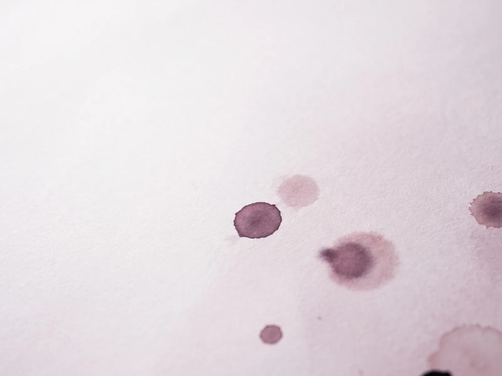
A port-wine stain — clinically, a port-wine birthmark (PWB) or capillary malformation — is a flat, pink, red, or purple mark present at birth, caused by a congenital malformation of the small dermal blood vessels. It affects roughly 3 in 1,000 newborns, is equally common across sexes and ethnicities, and never goes away on its own. Left untreated it tends to darken from pink toward purple and to thicken over decades, sometimes developing a bumpy or nodular surface in adulthood.
The treatment is laser, and the gold standard — by a wide margin — is the pulsed dye laser (PDL). This article explains why PDL is first-line, why treating early (including in infancy) changes the outcome, which lasers are used when PDL stalls, the Sturge-Weber syndrome screening that has to happen alongside any facial birthmark, and what laser realistically can and cannot achieve. It is educational and not a substitute for evaluation by a dermatologist or laser surgeon experienced in vascular birthmarks.
What a port-wine stain actually is
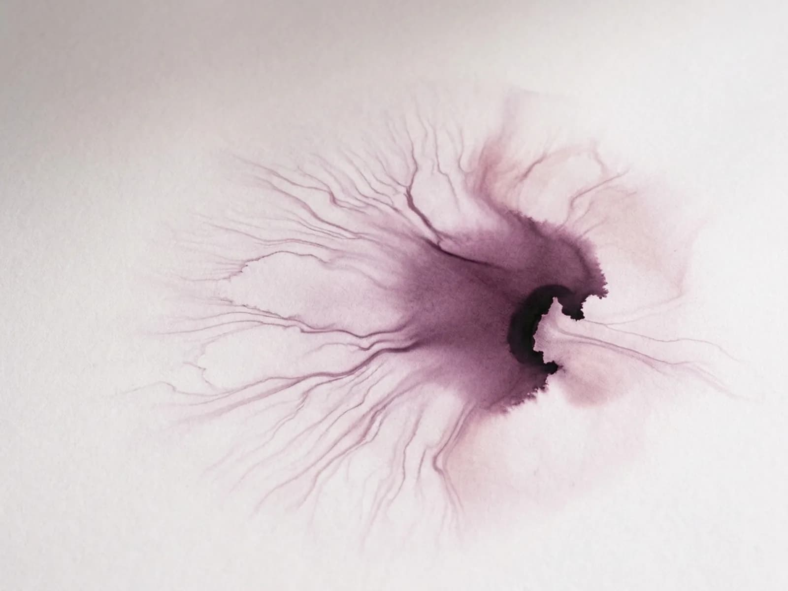
A PWB is not a collection of broken capillaries and not a hemangioma (a different lesion that grows and then shrinks). It is a static malformation: ectatic — abnormally widened — capillary-like vessels in the superficial dermis, lined by nerve cells that fail to regulate vessel tone, so the vessels stay dilated. The 2013 discovery that PWBs and Sturge-Weber syndrome are caused by a somatic mosaic mutation in the GNAQ gene (most commonly the p.R183Q substitution on chromosome 9q21) confirmed what clinicians had long suspected: the mark is laid down during embryonic development and is permanent without intervention.
That biology explains two things patients need to hear. First, the mark has a structural component that no cream touches. Second, it is progressive — the vessels widen further with age, the color deepens, and in many adults tissue overgrowth and nodularity develop. Early laser treatment is not only cosmetic; it can reduce that progression.
Not every red birthmark is a port-wine stain
A PWB is present and fully formed at birth, flat, and pink-to-red. It is distinct from two look-alikes parents should know about. An infantile hemangioma is usually not present (or is barely visible) at birth and instead grows over the first weeks to months before eventually involuting on its own — the opposite of a PWB's static-then-progressive course, and a completely different management pathway. Nevus simplex (the common "stork bite" or "angel kiss" on the nape, eyelid, or forehead) is a lighter pink patch that, unlike a PWB, fades substantially or completely in the first years of life. A dermatologist can usually distinguish these on exam; the distinction matters because a true PWB warrants laser consideration and SWS screening, while a nevus simplex typically does not.
The safety gate: Sturge-Weber syndrome screening
The most important step before any laser is taken is not about the laser. A facial port-wine birthmark, especially one on the forehead or in the distribution of the upper trigeminal nerve (V1), carries a risk of Sturge-Weber syndrome (SWS) — a congenital neurocutaneous disorder involving the skin, brain, and eye, with an estimated prevalence of roughly 1 in 20,000 to 1 in 50,000 live births. The same GNAQ mutation underlies both the birthmark and, when it affects the forebrain region, the leptomeningeal angioma and glaucoma that define SWS.
Forehead and upper-face PWBs are the highest-risk locations. Any infant with a facial PWB — particularly in the V1 (forehead/upper eyelid) distribution — should be evaluated for brain and eye involvement, typically with ophthalmology assessment for glaucoma (which can appear early) and, when indicated, neuroimaging. This workup happens in parallel with, not after, laser planning. Laser treats the skin; it does nothing for the brain or eye involvement of SWS, which need their own surveillance.
Pulsed dye laser — the gold standard and first-line treatment
The pulsed dye laser (585 nm, and now predominantly 595 nm) targets oxyhemoglobin in blood — the chromophore framework applied to vessels. The absorbed light heats and damages the ectatic capillary wall, the vessel is cleared by the body, and the visible color lightens. PDL has the longest efficacy and safety history of any device for PWB and is considered standard of care and first-line in the United States, including in infants.
Two findings have shaped modern practice:
- Treating in infancy is both safe and more effective. A landmark 2019 JAMA Dermatology study (Jeon, Bernstein, Geronemus and colleagues) showed pulsed dye laser treatment of PWBs in infancy could be performed without general anesthesia, and that earlier treatment improves lightening — younger skin has smaller, more superficial vessels and less established ectasia.
- Shorter intervals between sessions clear faster in infants. More recent research on infant PWS found that treating early and often — on shorter intervals rather than the traditional weeks-to-months spacing — accelerates clearance, which has pushed many pediatric laser centers toward tighter treatment spacing for babies.
- Multiple sessions are the rule, not the exception. Good clearance commonly takes roughly 8 to 10 treatments, and complete resolution is frequently not achieved. Patients and parents should expect a course, not a one-and-done procedure, and should understand that even well-treated PWBs can re-darken over years and need maintenance.
When PDL stalls: resistant, nodular, and hypertrophic lesions
PDL reaches only about the most superficial 1 mm of dermis, so it clears small surface vessels well but leaves deeper and larger vessels behind. This is why a PWB that responds beautifully at first often plateaus. For PDL-resistant, deeper, larger-vessel, or thickened/nodular PWBs, longer-wavelength devices are used because they penetrate deeper (roughly 50–75% deeper than PDL) with less scattering and less melanin absorption:
- Long-pulsed 1064 nm Nd:YAG reaches deeper, larger vessels and is used for hypertrophic and nodular PWB. It requires an experienced laser surgeon: even with cooling, the Nd:YAG can scar at or just above purpuric doses, so it is reserved for practitioners with the skill to titrate fluence.
- Long-pulsed 755 nm alexandrite is another near-infrared option for resistant and nodular lesions.
- 532 nm KTP (Nd:YAG, frequency-doubled) targets more superficial pigment/vessels and is used for finer residual vessels.
- Combination and sequential treatment — PDL followed by Nd:YAG, or a dual-wavelength (595 + 1064 nm) device — is used for mixed-vessel PWBs, treating different vessel sizes and depths in one session.
Adjuncts that are still emerging rather than standard: topical rapamycin (sirolimus) combined with PDL showed benefit in a phase II randomized trial for capillary malformations in SWS; HMME-PDT (hemoporfin photodynamic therapy), more commonly used in Asia (particularly China), is an alternative light-activated approach. These are not first-line in US practice but reflect an active research frontier.
Skin-of-color considerations
Port-wine stains occur in all skin types, and the Fitzpatrick phototype changes the risk calculus. In darker skin (Fitzpatrick IV–VI), the competing melanin absorption at 585/595 nm raises both the risk of post-inflammatory hyperpigmentation and the energy required to reach the vessel — a difficult tradeoff. Practical adjustments include lower starting fluence, more aggressive dynamic cooling, conservative test spots, longer intervals between sessions, and a relative preference for longer wavelengths (1064 nm) where deeper vessels need treating. Pigment changes after PDL are usually transient but can persist, and they should be discussed before the first session, not after.
Realistic outcomes and what laser cannot do
Laser lightens most PWBs, and early treatment improves both the degree of lightening and the chance of arresting progression. But expectations need calibration: complete clearance is the exception, not the rule. Most patients reach a meaningful but partial lightening after a multi-session course; many need maintenance treatments over the years as vessels slowly re-dilate; and thickened, nodular adult PWBs respond far less than flat pink infantile marks. Adverse effects to weigh include purpura (bruising, common and transient), swelling, blistering, pigment change (hypo- or hyperpigmentation), and — mainly with Nd:YAG — scarring. For an adult with an established nodular PWB, the realistic goal is softening and partial lightening, not a return to normal skin.
What it costs and how access works
PDL for a congenital PWB is sometimes covered by insurance, especially for children and for facial marks with functional or significant psychosocial impact, but coverage is inconsistent and often requires documentation and appeals. When self-pay, per-session pricing typically falls in the $300–$700 range depending on size, location, device, and practice, and a realistic course of 6–10+ sessions can run into the low thousands to mid-thousands. Pediatric vascular-anomaly centers at academic hospitals are often the best option for infants and children because they combine experienced laser surgeons, anesthesia support when truly needed, and the SWS workup under one roof. The number to compare is the total for the planned course, with the understanding that maintenance sessions may follow.
What to ask a provider — especially for parents
- Has my child been evaluated for Sturge-Weber syndrome — eye exam for glaucoma and neuroimaging if the mark is on the forehead (V1)? This is non-negotiable before or alongside laser.
- Are you starting with pulsed dye laser? It should be first-line.
- How many sessions should we expect, and how far apart? A credible answer is in the range of 8–10+ for meaningful clearance, spaced weeks to months apart.
- Can you treat without general anesthesia in an infant? Experienced pediatric laser centers often can.
- If PDL plateaus, what is the plan for deeper or resistant vessels (Nd:YAG, alexandrite)? And who is performing it — scarring risk rises with deeper lasers.
- Given my or my child's skin type, what is the pigment-change risk, and how is it minimized?
- What is the realistic endpoint — lightening, and how much — and will maintenance be needed?
A port-wine stain is permanent, but its color, progression, and psychosocial impact can almost always be improved with well-chosen, well-timed laser treatment. The patients and families with the best outcomes are those who get the SWS workup done, start PDL early with an experienced laser surgeon, and calibrate expectations around lightening and maintenance rather than erasure.
Sources
- JAMA Dermatology — Consensus Statement for the Management and Treatment of Port-Wine Birthmarks in Sturge-Weber Syndrome (PDL gold standard, infancy treatment, SWS screening): https://pmc.ncbi.nlm.nih.gov/articles/PMC8547264
- Pediatrics in Review — Port-wine Birthmarks: Update on Diagnosis, Risk Assessment for Sturge-Weber (V1/forehead risk, glaucoma, early laser): https://sturge-weber.org/file_download/inline/041b0d46-f550-4a20-a161-745ff57185cf
- PMC — The Pathogenesis of Port Wine Stain and Sturge-Weber Syndrome (GNAQ, MAPK/PI3K, anti-angiogenesis + PDL): https://pmc.ncbi.nlm.nih.gov/articles/PMC6539103
- Frontiers in Human Neuroscience — GNAQ mutations drive port wine birthmark-associated Sturge-Weber syndrome (p.R183Q, pathobiology): https://www.frontiersin.org/journals/human-neuroscience/articles/10.3389/fnhum.2022.1006027/full
- JAMA Dermatology — Pulsed Dye Laser Treatment of Port-Wine Stains in Infancy Without General Anesthesia (Jeon, Bernstein, Geronemus, 2019): https://jamanetwork.com/journals/jamadermatology/fullarticle/2730196
- Dermatology Times — Port wine stains clear faster with shorter pulsed dye laser treatment intervals (infant early-and-often protocol): https://www.dermatologytimes.com/view/port-wine-stains-clear-faster-shorter-pulsed-dye-laser-treatment-intervals
- British Medical Laser Association — Laser treatment guidelines for PWS in children (PDL, Nd:YAG, alexandrite for resistant lesions): https://bmla.co.uk/wp-content/uploads/Laser-treatment-guidelines-for-PWS-in-children.pdf
- UC Davis eScholarship — Light-based treatment of pediatric port-wine birthmarks (near-infrared lasers, session counts, Fitzpatrick): https://escholarship.org/content/qt7hh8g06g/qt7hh8g06g.pdf
- Vascular Birthmarks Foundation — Port Wine Stain patient information and GNAQ finding: https://birthmark.org/birthmark/port-wine-stain